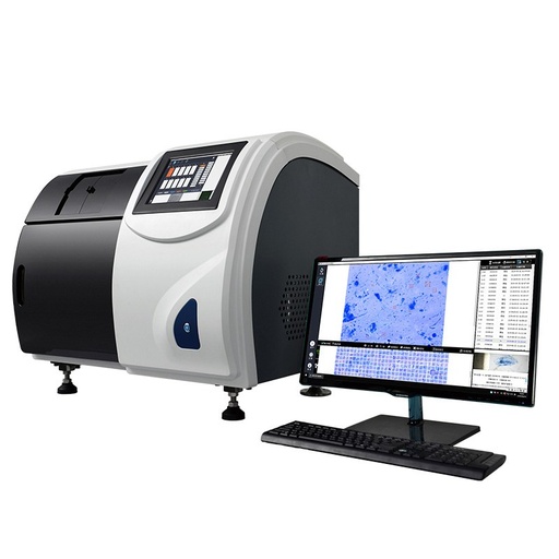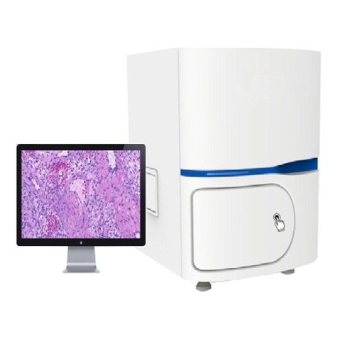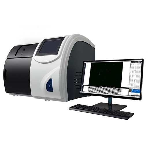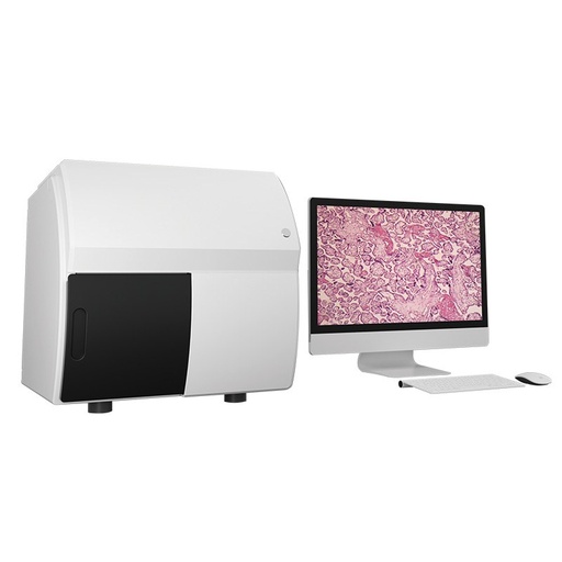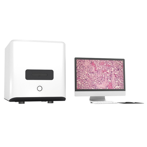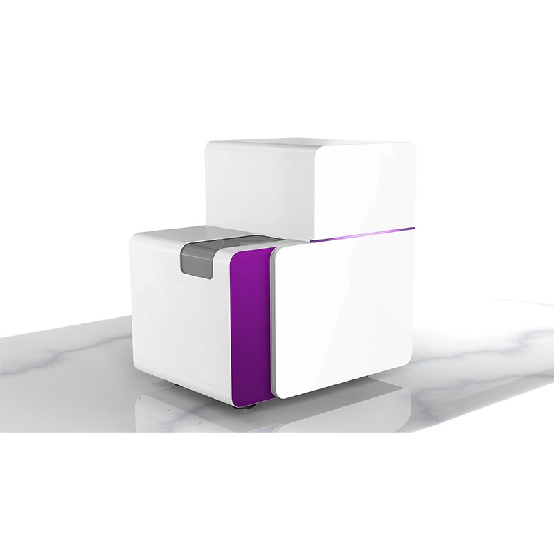
BestScope Scanpro5-30 Slide Scanning System
IntroductionThe Scanpro5 series slide scanning system adopts patented area scanning technology. It can scan slides with just one-click and the operation is simple. Through precise hardware control, stable operation is achieved. Through software and intelligent platform, system expansion can be realized, creating an intelligent diagnostic system and digital platform.
Model: Scanpro5-30 | Throughput: 30 | Objective: Plan apochromatic objective 20X, NA 0.8 | Scanning Method: Area scanning | Scanning Speed : 35s (20X scan) 50s (40X scan) | Dimensions: 621mm*632mm*439mm
Terms and Conditions
30-day money-back guarantee
Shipping: 2-3 Business Days
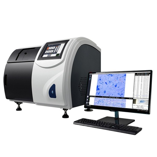
Scanpro5-30 Slide Scanning System
IntroductionThe Scanpro5 series slide scanning system adopts patented area scanning technology. It can scan slides with just one-click and the operation is simple. Through precise hardware control, stable operation is achieved. Through software and intelligent platform, system expansion can be realized, creating an intelligent diagnostic system and digital platform.
Key Details
| Model | Scanpro5-30 |
| Throughput | 30 |
| Objective | Plan apochromatic objective 20X, NA 0.8 |
| Scanning Method | Area scanning |
| Scanning Speed | 35s (20X scan) 50s (40X scan) |
| Dimensions | 621mm*632mm*439mm |
Feature
- Scanpro5-5
- Portable clinical-research all-in-one system
- High-definition imaging
- User-friendly operation
- Real-time viewing
- Scanpro5-60/120/180/240/300/360/420/480
- Expandable high-throughput intelligent scanning system
- Patented area scan technology, submicron panoramic imaging
- Original large-format area scan camera
- High-quality plan apochromatic objective, true color reproduction
- Scanpro5-30
- Smart mid-throughput scanning workstation
- Precision hardware control and area scanning for stability and clarity
- Power-off monitoring and smart reset to protect slides
Application
Automatic high-speed batch scanning
The digital slices are transmitted to the cloud for collaborative consultation
Combined with intelligent diagnostic software for auxiliary diagnosis
Specification
Model | Scanpro5-5 | Scanpro5-30 | Scanpro5-60/120/180/240/300/360/420/480 | ||
Throughput | 5 | 30 | 60/120/180/240/ 300/360/420/480 | ||
Scanning Method | Area scanning | ||||
Scanning Speed (15mm*15mm) | 35s (20X scan) 50s (40X scan) | ||||
Objective | Plan apochromatic objective 20X, NA 0.8 | ||||
Scanning Resolution | ≤0.24μm/pixel (20X scan) ≤0.12μm/pixel (40X scan) | ||||
Features | Supports multi-layer fusion scanning, supports slide label recognition, supports tissue contour recognition, supports slide path monitoring, supports slide upload and QR code sharing | ||||
Output Image Format | TMAP/SVS/TIFF, etc. | ||||
Dimensions | 565mm*425mm*563mm | 621mm*632mm*439mm | 896mm*635.5mm*715mm | ||
Software & Platform
1.Smart pathology cloud platform (Optional)
The intelligent smart pathology cloud platform deeply integrates cloud computing, artificial intelligence, and pathology to create a comprehensive information platform combining diagnosis, collaboration, management and education.
- Collaboration: Remote consultations pool expert wisdom
- Efficiency: Process optimization shortens report time
- Precision: AI assistance reduces missed and misdiagnoses
- Standardization: Quality control standards ensure diagnostic accuracy
- Sharing: Case sharing fosterscollective progress
It addresses regional pathology challenges by leveraging an integrated platform to implement tiered healthcare
- Deep integration of AI for accurate diagnosis and full-process quality control.
The platform deeply integrates artificial intelligence into diagnosis and quality control, providing pathologists with objective decision-making support to enhance diagnostic accuracy and efficiency, enabling refined management of operations and ensuring robust diagnostic quality.
- Empowering medical alliances and grassrootsunits to promote resource collaboration and sharing.
By establishing a cloud-based pathology diagnosis center, an efficient model of grassroots testing, superior diagnosis can be achieved. This effectively alleviates the shortage of pathologists and limited diagnostic capabilities at the grassroots level, while optimizing the allocation of regional medical resources, making it a key tool for implementing tiered healthcare.
- Building a data hub to drive clinical research and service innovation
Promotes mutual recognition and sharing of test results with regions, enables centralized supervision of regional healthcare big data, and allows patients to conveniently access reports via mobile devices, thereby improving the healthcare experience.
Regional integrated platform architecture diagram
2.Cervical cell-Image processing software (Optional)
LSIL and above, sensitivity ≥99%, specificity ≥80%. Using standard slide samples from the pathology departments of 17 hospitals, and after excluding abnormal samples, the sensitivity for LSIL and above was 100% and the specificity was 81% in 100,612 cases.
- Combined dual screening: One stain, two reports (TCT + DNA)
- Microbial infection alert: Fungus, actinomyces, clue cells, etc.
- Strong robustness: Compatible with: membrane-based, sedimentation, centrifugation, etc.
Primary Screening Classification Table | ||
Level 1 Classification | Level 2 Classification | Level 3 Classification |
Negative | No Intraepithelial Lesion | Metaplasia, hemolysis, repair, coagulation, post-review |
Positive | Suspected epithelial cancer cell density | AGC-NOS, AGC-FN, AIS |
Squamous epithelial cell abnormalities | ASC-US, ACS-H, LSIL, HSIL, SCC | |
Advantages of combined screening:
- Comprehensive analysis: DNA ploidy + TBS2014 classification.
- Microbial infection detection and immune response display.
- Direct reanalysis and atypical bacterial testing.
- Rapid Localization and online review of suspicious cell fields of view.
- Slide satisfaction evaluation based on TBS standards.
- Support for scanning quality assessment.
Cervical cytology TBS intelligent analysis
Cervical cytology DNA ploidy quantitative intelligent detection
3.Histopathology-Image processing software (Optional)
It covers high-incidence cancer types in various tissues, including stomach, intestine, lung, etc. It can complete slide analysis in as fast as 1 minute, reducing diagnosis time to within 6 hours. It is Intelligent and efficient and supports adaptation to multiple devices. The private deployment ensures data security.
It supports intelligent tumor typing and benign / malignant classification:
Functional interface features:
- Quick positioning and online review of suspicious cell fields of view
- Real-time interpretation with precise insights into lesions
- Flexible switching between multiple magnifications to meet various slide reading needs
- Human-machine collaborative comprehensive diagnostic decision-making
- Multiple visualization methods for results, enabling rapid localization of lesion areas
Heatmap visualization of tissue lesion contours
Dimension
Unit: mm