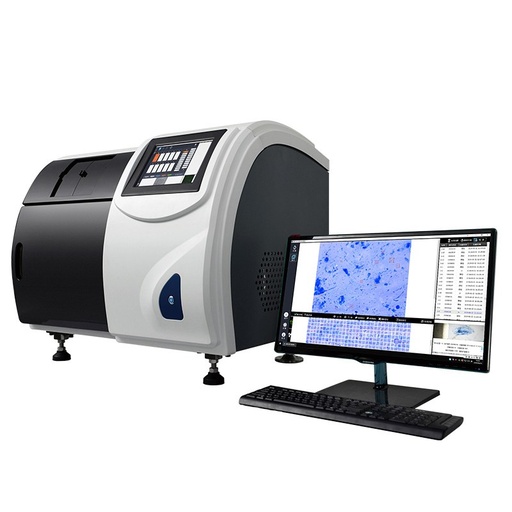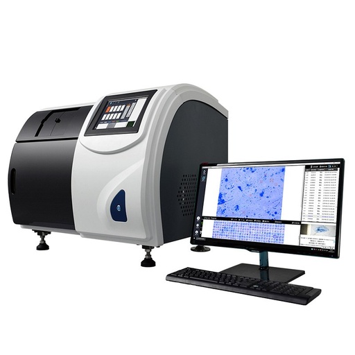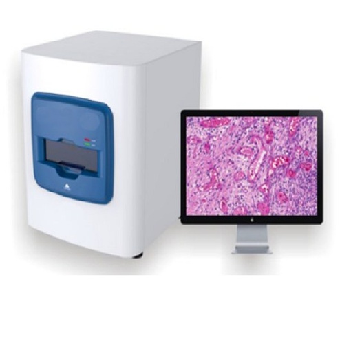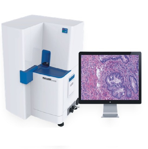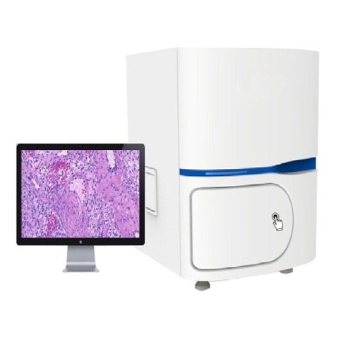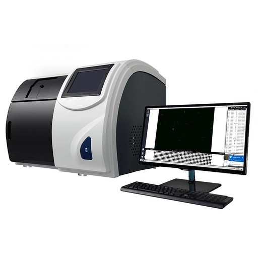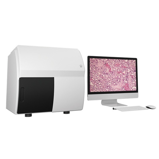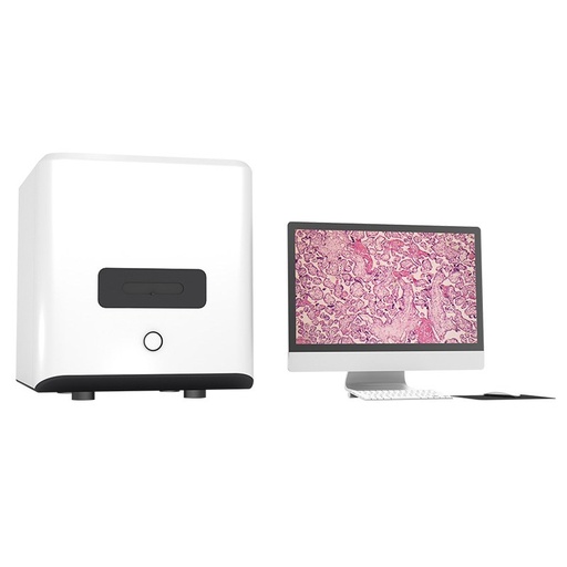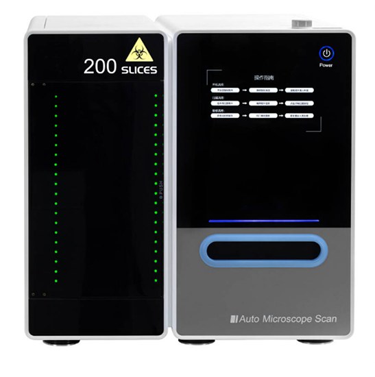
BestScope Scanpro4 Whole Slide Imaging Scanner
IntroductionIn the research process of life science, microscopic observation is an indispensable technique. But traditional microscopic observation is limited by time, region, personnel and equipment. Specimens must be observed in the laboratory by professionals using a microscope device. The Whole Slide Imaging (WSI) technology is an organic combination of modern digital systems and traditional optical magnification devices. After digital…
Model: Scanpro4 | Illuminator: LED | Objective: 10X, 20X, 40X, 60X and 100X | Panoramic Camera: 5 mega-pixels | Power Consumption: 220W | Power Rating: Input: 220V AC, 50Hz, 1A
Terms and Conditions
30-day money-back guarantee
Shipping: 2-3 Business Days
Scanpro4 Whole Slide Imaging Scanner
IntroductionIn the research process of life science, microscopic observation is an indispensable technique. But traditional microscopic observation is limited by time, region, personnel and equipment. Specimens must be observed in the laboratory by professionals using a microscope device. The Whole Slide Imaging (WSI) technology is an organic combination of modern digital systems and traditional optical magnification devices. After digital imaging of specimens through this technology, researches can perform digital reading at any location, realizing a revolution in digital imaging and reading.
Key Details
| Model | Scanpro4 |
| Illuminator | LED |
| Objective | 10X, 20X, 40X, 60X and 100X |
| Panoramic Camera | 5 mega-pixels |
| Power Consumption | 220W |
| Power Rating | Input: 220V AC, 50Hz, 1A |
Feature
Bright field & fluorescence imaging methods
The product has both bright field and fluorescence imaging methods, allowing researchers to switch freely in and application.
Support standard slides and various custom size slides
Multi objective lens system
Scanpro4 series support configuration of multiple objective lenses, up to 3. It is convenient for researchers to observe specimens from low magnification to high magnification, or even oil lens, supporting various application scenarios and projects.
Automatic oil filling
The automatic oil dripping system can meet the needs of researchers for oil lens observation. The automatic oil dripping system eliminates the trouble of researchers frequently oiling the objective lens
Multi feeding mechanism
The feeding mechanism is designed to be 5 pieces, 50 pieces, 100 pieces, 200 pieces, (it can be customized to upgrade to 400 pieces, 600 pieces, of higher throughput) Each tray can hold up to 5 standard slides, and the tray-type design ensures the safety of specimens, without broke or stuck. The label recognition of slide specimens can be realized before the specimen is scanned, and barcode/QR code is supported. Panoramic image acquisition and algorithms before scanning can help researchers locate the scanning area more quickly, or manually set scanning parameters to meet individual needs.
Efficient and user-friendly operation
The operation interface and image reading and observation interface are simple and easy to use. The concise working interface provides the greatest convenience for researchers. Digital slice reading software (local/remote).
The software can be accessed through local or network storage to view microscopic digital slices, and can also view uploaded microscopic digital slice images through internet connection or local area network connection. Support image annotation, measurement and other functions.
Application
Microorganism
Microbial smears are commonly used for clinical inspection, teaching and scientific research. Microorganisms are analyzed by observing the microstructure, quantity and dynamics of microorganisms such as bacteria and fungi. The Scanpro4 slide scanner can convert microbial smear specimens into digital slides, enabling digital reading at multiple terminals.
Gram-negative bacteria Escherichia coli
Nissl staining
Auramine O staining
Pathology
In histopathological and cytopathological studies, the ability to assess cell morphology and analyze two objects (localization) in close proximity or overlapping with each other is critical. Based on the accumulation of optical technology for many years, the optical system of Scanpro4 slide scanner can make the target cells clearer. The cells and tissues can be seen at a glance, and the pathological section can be comprehensively evaluated.
FISH fluorescence
Immunofluorescence technology is often used in disease diagnosis and drug development to analyze samples. Scanpro4 slide scanner can detect multiple molecular targets in samples, gathering information from multiple molecular targets onto a digital slide through image fusion. It provides a powerful help for the researchers to analyze the results.
Multicolor fluorescence fusion image
Blood
Blood is one of the most important tissues in the human body. The changes of white blood cells, platelets and red blood cells in the blood reflect the health and metabolic level of the human body. In clinical inspection work, blood cell morphological examination is a basic but very important inspection item. Scanpro4 slide scanner can convert blood cell smears into digital slides, and provide high-definition and comprehensive blood cell images for inspection personnel on the reading software for diagnosis.
Blood cell morphology analysis
Specification
Item | Specification |
Observable Sample | Slides with/without standard coverslips |
Slide Size | Standard slide trays (L*W*H, 5 slides): 75mm-76.5mm (2.95in-3in), 25mm-26.5mm (0.98in-1in), 0.9mm-1.2mm (0.04in inches-0.05 inches) |
Optional slide tray 1 (L*W*H, 2 slides): 75mm-76.5mm (2.95in-3in), 51mm to 53mm (2 in-2.09in), 0.9mm-1.2mm (0.04 inches-0.05 inches) | |
Optional slide tray 2 (L*W*H, 1 slide): 100mm-102mm (3.94in-4.02in), 75mm-76.5mm (2.95in-3in), 0.9-1.2mm (0.04 inches-0.05 inches) | |
Observation Method | Bright field |
Fluorescence | |
Illuminator | Built-in transmitted light Kohler illumination, high intensity and high color rendering LED (up to 50,000 hours lifetime) |
Objective Lens | Compatible objectives: 10X, 20X, 40X, 60X and 100X. 3-position motorized turret (includes select oil immersion) |
Automatic oil distributor | |
Stage | Automatically controlled motorized XY stage |
Focusing | Automatically controlled autofocus with support for focused topographic maps and real-time autofocus |
Scan Camera | Color camera, 1.1” CMOS, 4.5um*4.5um pixels, 7 mega-pixels, high sensitivity, high resolution |
Monochrome camera, 1.1” CMOS, 4.5um*4.5um pixels, 7 mega-pixels, high sensitivity, high resolution | |
Panoramic Camera | Color Camera, 5 mega-pixels |
Capacity | 5 slides: 1 slide tray, up to 5 slides, can be upgraded to multi-tray feeding mechanism |
100/200 slides: up to 40 slides trays, up to 200 slides | |
400/600 pieces can be customized | |
Pixel Resolution | 10X (NA 0.4): 0.672 um/pixel |
20X (NA 0.8): 0.335um/pixel | |
40X (NA 0.95): 0.168um/pixel | |
60X (NA 1.4): 0.112um/pixel | |
100X (NA 1.35): 0.067um/pixel | |
Scanning Time | Bright field is less than 60 seconds (20X objective lens, scanning area 15mm*15mm) |
Software | Auto barcode reading, auto focus topographic map, auto scan, auto stitching, pause and resume scanning, Z-stack imaging, multiple image formats (BMP, JPEG and TIFF), simultaneous multi-image display, stepless zoom, upload while scanning slide browsing, annotation, screen capture |
Fluorescence | Equipped with professional LED fluorescent illuminator, motorized 4-hole fluorescent reflected lens turntable, motorized filter wheel, fluorescent light source |
Oil component | Long life, maintenance-free, 5 million times |
Weight | Slide scanner of 5 slides: 40kg, slide scanner of 200 slides: 70kg. 1 slide tray: 0.1 kg |
Operating Environment | Temperature: 10°C-30°C. Humidity: up to 80% (non-condensing) |
Power Consumption | 220W |
Power Rating | Input: 220V AC, 50Hz, 1A |
Sample Image
Immunofluorescence slide
Plant slide
Tissue sections slide
Chromosome karyotype slide
Dimension
Unit: mm
Specifications for BestScope Scanpro4 Whole Slide Imaging Scanner
| Manufacturer | BestScope |